10+ Tfcc Injury Mri Radiology Pics - Hi buddy 4k Free Wall, In the article that you are reading this time with the title 10+ Tfcc Injury Mri Radiology Pics, we have prepared this article well for you to read and take the information in it. hopefully the content of the post what we write you can understand. all right, have a nice reading.
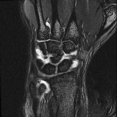
10+ Tfcc Injury Mri Radiology Pics. The triangular fibrocartilage complex (tfcc) is a complex structure that is a major contributor to the stability of the wrist. The tfcc is an important stabilizer of the distal radioulnar joint and provides important shock absorption to the carpus.
Peripheral Tears of the TFCC: Arthroscopic Diagnosis and ... from musculoskeletalkey.com The triangular fibrocartilage complex (tfcc) is a complex structure that is a major contributor to the stability of the wrist. Review this list very carefully. Arthroscopy is the diagnostic gold standard.
Magnetic resonance imaging (mri) is a proven, established imaging modality for the detection, evaluation, staging, and follow up of disorders of the wrist.
Triangular fibrocartilage complex injury exercises or tfcc injury exercises. The subsequent loss of alignment with time produces the mri signal. Palmer class 1b tfcc injury. The triangular fibrocartilage complex (tfcc) is a complex structure that is a major contributor to the stability of the wrist.
Thus the article 10+ Tfcc Injury Mri Radiology Pics
That's the article 10+ Tfcc Injury Mri Radiology Pics this time, hopefully it can be of benefit to all of you. well, see you in another article post.
You are now reading the article 10+ Tfcc Injury Mri Radiology Pics with the link address https://hooveranniversaryeditionbuyonline.blogspot.com/2021/09/10-tfcc-injury-mri-radiology-pics.html
Share this post
0 Response to "10+ Tfcc Injury Mri Radiology Pics"
0 Response to "10+ Tfcc Injury Mri Radiology Pics"
Post a Comment